Seminar 46 - Images
Case 2
Dr. Gerald Berry
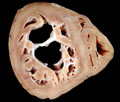 |
Fig. 1 Histiocytoid Cardiomyopathy |
Case 4
Dr. Ira Bleiweiss
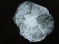 |
Fig. 1 |
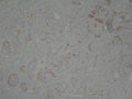 |
Fig. 2 |
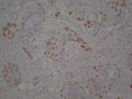 |
Fig. 3 |
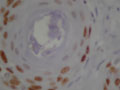 |
Fig. 4 |
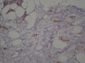 |
Fig. 5 |
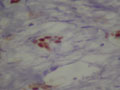 |
Fig. 6 |
Case 6
Dr. Thomas Colby
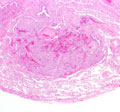 |
Fig. 1 The case presented (AMR 46, T. Colby's case) shows a 3 mm focus of metastatic meningioma surrounding a bronchovascular bundle suggesting lymphangitic growth. |
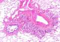 |
Fig. 2 Another case of metastatic, histologically benign meningioma showing fairly convincing lymphangitic growth along a bronchovascular bundle. This was a microscopic focus identified adjacent to a grossly identifiable nodule. |
Case 9
Dr. Masaharu Fukunaga
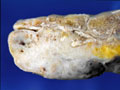 |
Fig. 1 |
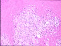 |
Fig. 2 |
Case 13
Dr. Elizabeth Montgomery
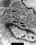 |
Fig. 1 |
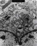 |
Fig. 2 |
Case 16
Dr. Dominic Spagnolo
 |
Fig. 1 |
 |
Fig. 2 |
 |
Fig. 3 |
 |
Fig. 4 |
 |
Fig. 5 |
Last update: August 7, 2023
Copyright © 2004-2024 AMR International Pathology Slide Seminar Club