Bisceglia, Case 2
Clinical history :
A 3.5-cm sized nodular lesion was removed from the right parotid region in a very young girl aged 13. The lesion had appeared 2 months before. However the past medical history of this young patient was remarkable for several previous surgical excisions of cutaneous benign fibrous histiocytoma with repeated recurrences in the head and neck area.
 Figure 1
|
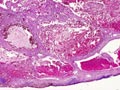 Figure 2
|
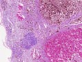 Figure 3
|
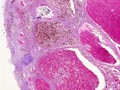 Figure 4
|
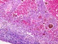 Figure 5
|
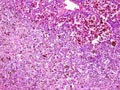 Figure 6
|
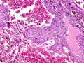 Figure 7
|
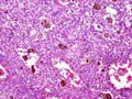 Figure 8
|
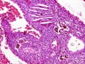 Figure 9
|
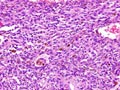 Figure 10
|
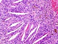 Figure 11
|
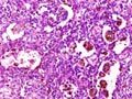 Figure 12
|
Last update: July 23th, 2005
Copyright © 2004-2005 Biopticka laborator s.r.o.
Copyright © 2004-2005 Biopticka laborator s.r.o.