Colby, Case 2
Clinical history :
A 33-year-old woman with tuberous sclerosis presented with renal failure and abdominal enlargement. She was known to have bilateral angiomyolipomas, had previously had pneumothoraces, and had documented pulmonary lymphangioleiomyomatosis. Two years prior to presentation she was placed on oral contraceptives, and noted increase in size of her renal masses. An umbilical biopsy showed endometriosis. Clinically she had adenoma sebaceum of the face and trunk, but she was not retarded, and had no CNS lesions. At laparotomy both kidneys had cystic masses that were resected. She also had a hysterectomy and bilateral salpingo-oophorectomy. Endometriosis was documented in the pelvis. Lymphatics in the pelvic retroperitoneum were abnormal. After surgery she was taken off hormonal therapy, placed on a low protein diet, and was stable with minimal shortness of breath three months after surgery.
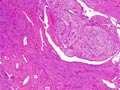 Figure 1
|
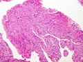 Figure 2
|
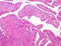 Figure 3
|
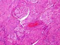 Figure 4
|
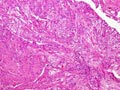 Figure 5
|
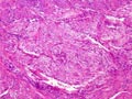 Figure 6
|
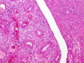 Figure 7
|
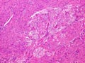 Figure 8
|
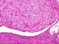 Figure 9
|
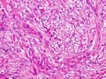 Figure 10
|
Copyright © 2004-2005 Biopticka laborator s.r.o.