Cooper, Case 2
Clinical history :
A 93-year-old man presented with progressive left-sided weakness and paralysis. A large soft tissue mass with bone destruction involving T3 was detected on CT scan of the thoracic spine. There was considerable extension of tumor into the spinal canal with marked canal compression. Surgery was undertaken to decompress the pressure on the spinal cord with several fragments (largest 1.2 x 0.5 cm) of tumor tissue being submitted for histological examination.
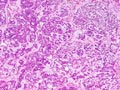 Figure 1
|
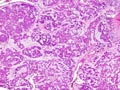 Figure 2
|
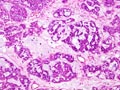 Figure 3
|
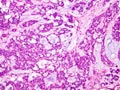 Figure 4
|
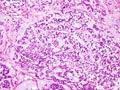 Figure 5
|
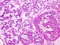 Figure 6
|
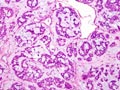 Figure 7
|
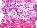 Figure 8
|
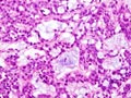 Figure 9
|
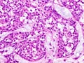 Figure 10
|
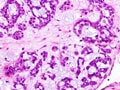 Figure 11
|
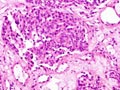 Figure 12
|
Last update: July 23th, 2005
Copyright © 2004-2005 Biopticka laborator s.r.o.
Copyright © 2004-2005 Biopticka laborator s.r.o.