Falconieri, Case 2
Clinical history :
A 39 year-old man complains of abnormal weight gain, headache, bitemporal hemianopsia. A MRI scan reveals a solid mass of the third ventricle. Craniotomy is performed and several, grey tan tissue fragments are resected.
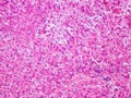 Figure 1
|
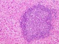 Figure 2
|
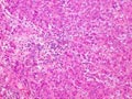 Figure 3
|
 Figure 4
|
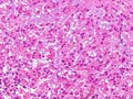 Figure 5
|
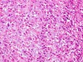 Figure 6
|
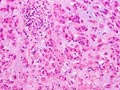 Figure 7
|
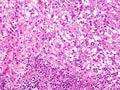 Figure 8
|
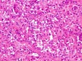 Figure 9
|
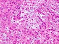 Figure 10
|
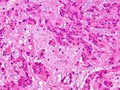 Figure 11
|
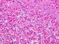 Figure 12
|
Last update: July 23th, 2005
Copyright © 2004-2005 Biopticka laborator s.r.o.
Copyright © 2004-2005 Biopticka laborator s.r.o.