Fukunaga, Case 1
Clinical history :
Three years prior to hospitalization, a 52-year-old Japanese, gravida 2, pare 2, woman noted a small mass in the vulva. The mass enlarged slowly. Physical examination revealed a 3 x 2.5 cm, nonfixed, nontender, rubbery, well-defined mass in the left labium majus. It did not involve the overlying skin. No inguinal lymph nodes were palpable. Laboratory date was unremarkable. The initial diagnosis of a punch biopsy was "poorly differentiated adenocarcinoma". Plain X-rays and computed tomography scan showed that there was no mass in the lung and liver. Wide excision of the vulva, total abdominal hysterectomy, bilateral salpingo-oophorectomy and the pelvic and inguinal lymph node dissection were performed because clinically the primary site was not known. Extensive clinical examination failed to find other possible primary site. The patient was well and free of disease at 25 months.
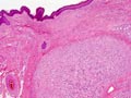 Figure 1
|
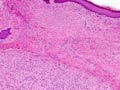 Figure 2
|
 Figure 3
|
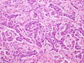 Figure 4
|
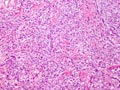 Figure 5
|
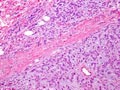 Figure 6
|
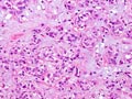 Figure 7
|
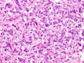 Figure 8
|
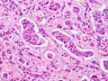 Figure 9
|
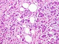 Figure 10
|
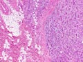 Figure 11
|
Copyright © 2004-2005 Biopticka laborator s.r.o.