Fukunaga, Case 2
Clinical history :
A 51-year-old, gravida 2, para 2, Japanese woman presented with left lower abdominal pain. Physical, computed tomography scan and ultrasound examinations indicated a cystic ovarian tumor. Laboratory data showed no abnormalities. Because of the risk of left-side ovarian cancer, abdominal total hysterectomy and bilateral salpingo-oophorectomy was performed. Intraoperatively, a subserosal, sessile polypoid mass with multiple cysts in the anterior fundus of the uterus was found, and the ovaries and tubes were unremarkable. The subsequent course has been uneventful for 24 months.
 Figure 1
|
 Figure 2
|
 Figure 3
|
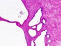 Figure 4
|
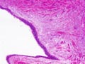 Figure 5
|
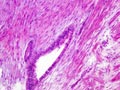 Figure 6
|
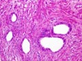 Figure 7
|
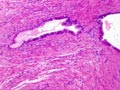 Figure 8
|
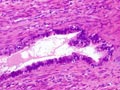 Figure 9
|
 Figure 10
|
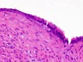 Figure 11
|
Last update: July 23th, 2005
Copyright © 2004-2005 Biopticka laborator s.r.o.
Copyright © 2004-2005 Biopticka laborator s.r.o.