Fukunaga, Case 3
Clinical history :
A 60-year-old man presented in October 1989 with a several month history of an elevated skin lesion measuring 1 cm in base of the second toe of the sole of the right foot. Biopsy, a wide excision and a skin graft were performed and the lesion was diagnosed as malignant melanoma, Clark level, II. The margins were free from the lesion. The tumor recurred in September 2003 and the patient underwent an amputation of the distal half of the left foot in February 2004. Physical examinations and a computed tomography scan revealed no other primary site and showed no metastatic lesions. No lymphoadenopathy was found. The patient has been well without disease for 10 months after the amputation.The distributed slide is taken from the recurrent tumor.
 Figure 1
|
 Figure 2
|
 Figure 3
|
 Figure 4
|
 Figure 5
|
 Figure 6
|
 Figure 7
|
 Figure 8
|
 Figure 9
|
 Figure 10
|
 Figure 11
|
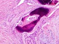 Figure 12
|
 Figure 13
|
 Figure 14
|
 Figure 15
|
 Figure 16
|
 Figure 17
|
Copyright © 2004-2005 Biopticka laborator s.r.o.