Mentzel, Case 1
Clinical history :
A 34-year-old female patient developed a slowly growing deep seated neoplasm in her right thigh. No previous trauma was reported. MRI-scans showed a well-circumscribed, intramuscular lesion. After an initial biopsy the neoplasm was completely excised. Grossly, a 10.9 cm measuring nodular lesion with central necrosis and haemorrhage was described. There is no sign of recurrence at 54 months.
 Figure 1
|
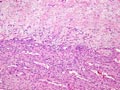 Figure 2
|
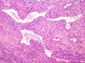 Figure 3
|
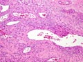 Figure 4
|
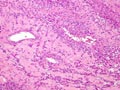 Figure 5
|
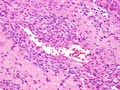 Figure 6
|
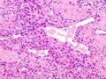 Figure 7
|
 Figure 8
|
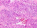 Figure 9
|
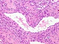 Figure 10
|
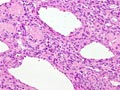 Figure 11
|
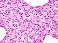 Figure 12
|
Last update: July 23th, 2005
Copyright © 2004-2005 Biopticka laborator s.r.o.
Copyright © 2004-2005 Biopticka laborator s.r.o.