Mentzel, Case 3
Clinical history :
In March 2003 a 54-year-old male patient presented with a long-stainding (more than 5 years), slowly growing lesion on his left palm. Clinically, an exophytic neoplasm measuring 9 x 9 cm was seen, however the patient still refused excision. Magnetic resonance imaging showed changes compatible with a lipomatous lesion. After a diagnostic biopsy in October 2003, the neoplasm was completely and with tumour free margins excised. Intraoperative a nodular, well-circumscribed as well as encapsulated neoplasm was noted. There is no sign of recurrence at 8 months.
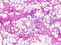 Figure 1
|
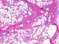 Figure 2
|
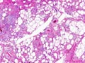 Figure 3
|
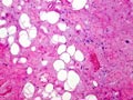 Figure 4
|
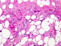 Figure 5
|
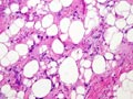 Figure 6
|
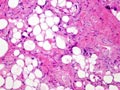 Figure 7
|
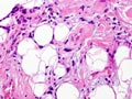 Figure 8
|
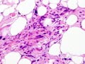 Figure 9
|
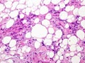 Figure 10
|
Last update: July 23th, 2005
Copyright © 2004-2005 Biopticka laborator s.r.o.
Copyright © 2004-2005 Biopticka laborator s.r.o.