Michal, Case 2
Clinical history :
Patient was 35-year-old man, who has gradually enlarged mass 4 x 5 x 5 cm in size in left testis. The patient was aware of the tumor since his age of 10. The testis was elastic and painless. Contralateral testicle was normal size and consistency. No enlarged lymph nodes or metastases were revealed by ultrasonography and computed tomography. Serum levels of alpha-fetoprotein, beta-HCG and CEA were normal. Left orchiectomy was performed. Grossly, the testis was replaced by uniform whitish soft tissue. The tumor was round-shaped, well circumscribed with thin capsule. Karyotype of the patient was normal (46XY). Eleven years after the excision, the patient is healthy and without signs of recurrence and metastases.
 Figure 1
|
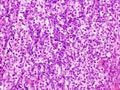 Figure 2
|
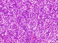 Figure 3
|
 Figure 4
|
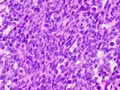 Figure 5
|
Last update: July 23th, 2005
Copyright © 2004-2005 Biopticka laborator s.r.o.
Copyright © 2004-2005 Biopticka laborator s.r.o.