Michal, Case 3
Clinical history :
The patient was a 46-year-old man who underwent a surgical excision of a sigmoid colon with an "inflammatory pseudotumor". Precise size of the lesion was not known although the lesion was described as a "very large" tumor of white fibrous appearance removed in many pieces weighing altogether 450 grams. In one of the excised tissue fragments we found an iron pin encased by the tumorous tissue.
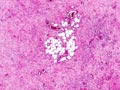 Figure 1
|
 Figure 2
|
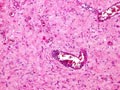 Figure 3
|
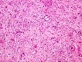 Figure 4
|
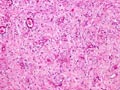 Figure 5
|
 Figure 6
|
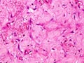 Figure 7
|
Last update: July 23th, 2005
Copyright © 2004-2005 Biopticka laborator s.r.o.
Copyright © 2004-2005 Biopticka laborator s.r.o.