Wakely, Case 2
Clinical history :
A 65-year-old man was admitted to the hospital with presumed acute prostatitis. A pelvic ultrasound showed an incidental 3.5 x 3-cm, solid, enhancing mass arising from the lower pole of the left kidney. There was no evidence of retroperitoneal adenopathy, renal vein invasion, local extension, or visceral metastases. A partial nephrectomy was performed.
 Figure 1
|
 Figure 2
|
 Figure 3
|
 Figure 4
|
 Figure 5
|
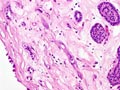 Figure 6
|
 Figure 7
|
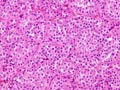 Figure 8
|
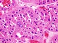 Figure 9
|
Last update: July 23th, 2005
Copyright © 2004-2005 Biopticka laborator s.r.o.
Copyright © 2004-2005 Biopticka laborator s.r.o.