Archive >> Archive of Images >> Seminar 34
Seminar 34 - Images
Case 4
Fig. 1
|

Fig. 2
|
Fig. 3
|
Case 5
The following pictures illustrating the well-differentiated component of the tumor were sent in a second time.
Fig. 1
|
Fig. 2
|
Case 8
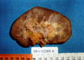
Fig. 1
|
Case 10
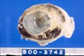
Fig. 1
|
Case 12
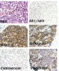
Fig. 1
|
Case 19
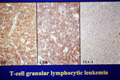
Fig. 1
|
Fig. 2
|
Fig. 3
|
Case 20
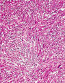
Fig. 1 - H&E
|
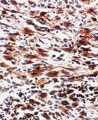
Fig. 2 - CD68
|
Last update: July 09, 2026
Copyright © 2004-2026 AMR International Pathology Slide Seminar Club